The stronger the better?
Ultra-high-field MRI has finally found its way into clinical practice. Despite large efforts in the ultra-high-field research, its full potential has yet to be realized.

At the end of August, the approval for the first ultra-high-field MRI scanner to be used in clinical practice in Europe was won and in October, the FDA cleared the first 7T MRI device for clinical use. Even though MRI scanners of such strength have been used in research hospitals for years, its current approval shows signs that ultra-high-field MRI will be integrated into more routine applications in the future.
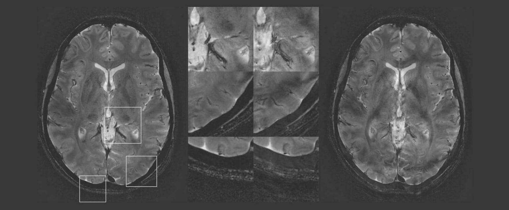
In his recent paper “Imaging at ultrahigh magnetic fields” in Neuroimage, Kamil Ugurbil took a closer look at the history and challenges of imaging at ultra-high-field and referred back to a time where a 1.5T scanner was considered “high-field”. With the introduction of 3T, later 7T and higher magnetic field platforms considerable advantages in signal-to-noise ratio were achieved. Ultra-high-field has proven to be specifically advantageous in the areas of brain function and anatomy, where it has brought fMRI to submillimeter resolution in the whole brain. It also provides exquisite anatomical detail in many organ systems of the human body boosting biomedical research and clinical diagnosis.
However, along with several advantages, ultra-high-field also brings numerous challenges. For example, fMRI relies on very fast image acquisition strategies that suffer from magnetic field inhomogeneities, which cause distortions, blurring and signal loss. Additionally, imaging the human brain tissue can be challenging due to large magnetic field inhomogeneities that increase with increasing field strengths. Moreover, physiological noise sources become more pronounced with stronger fields.
Together, such obstacles form a major challenge in ultra-high-field imaging which may impact the sensitivity and specificity of clinical diagnoses and research. Further improvements and research are thus crucial to ensure the efficient demployment of the 7T and potentially higher field MR scanners into the clinical environment where short scanning times are of essence.
Such practical implementation questions may make us wonder if the stronger magnetic fields are truly superior? It is clear that before higher magnetic field platforms will pave the way for human imaging with increasing biological information, capabilities to enable a robust and efficient use of such systems have to be acquired. Exciting times ahead!
The article has been updated after the press announcement by the FDA.