fMRI
Data quality in fMRI directly impacts your ability to detect neurological activation. Images reconstructed with the knowledge of field encoding dynamics have reduced noise and artifacts from scanner and physiologic sources, providing images with increased SNR and geometric accuracy for more powerful analyses.
fMRI is probing fundamental questions in human cognition and is helping to improve the lives of those with neurological diseases. Despite its prevalence in research and expanding use in the clinic, fMRI remains a challenging imaging technique due to it’s dependence upon accurate image encoding, geometric consistency, and high SNR.
fMRI records neurological activations, such as response to stimuli, as changes in image intensity. These intensity changes are small compared to noise inherent in the image and can be obscured by subtle image encoding errors from the scanner and physiological sources. Identifying neurological activations in group analyses requires images with high temporal SNR to ensure statistical confidence and accurate geometry to correctly localize the activation.
Accurately capturing image encoding improves image geometry, reduces ghosting, and improves SNR in fMRI imaging. Explore the next paragraphs to see how field monitoring could improve your fMRI acquisitions!

Take control of single-image and temporal SNR in fMRI
 |
Image SNR is critical to the detection of neurologic activations in fMRI. Image SNR is directly impacted by gradient performance. Pre-programmed gradient trajectories are used to encode the image during acquisition. Subtle differences between the programmed waveform and the output waveform cause undesired phase accrual and image artifacts, including ghosting and loss of SNR.
The best analyses require consistent image quality throughout the entire imaging session. Lengthy scan sessions and using gradients at a high duty cycle are common in fMRI and can induce frequency drift and gradient heating. Both of which can degrade the temporal SNR of fMRI acquisitions and this results in the reduced ability to detect functional activation of brain regions.
Skope field monitoring solutions capture the changing dynamics including field drift and gradient heating (left/right in the image above) to reconstruct images without distortions, allowing functional activation to be strongly detected.
Ensure accurate image geometry for each subject
Detecting group and population differences in neuroscience studies requires fMRI data to have correct image geometry for each subject.
Image geometry is impacted by various factors, including the accuracy of image encoding. The same subtle variations that impact SNR can also distort image geometry.
Skope field monitoring solutions measure the actual image encoding trajectory and reconstruct images accounting for field fluctuations which distort image geometry.
Take fMRI to higher resolution at ultra-high field
Ultra-high field (7T +) holds multiple promises for improving fMRI research. Faster relaxation times mean potentially greater image contrast, allowing better identification of activations; increased signal means higher SNR for the same resolution or increased resolution with SNR comparable to 3T. These promises, however, come with challenges. In fact, increased resolution means that smaller movements outside the field of view can degrade image quality.
Image reconstructions informed by field monitoring can help account for these movements, improving images and unlocking new applications for your research at ultra-high field.
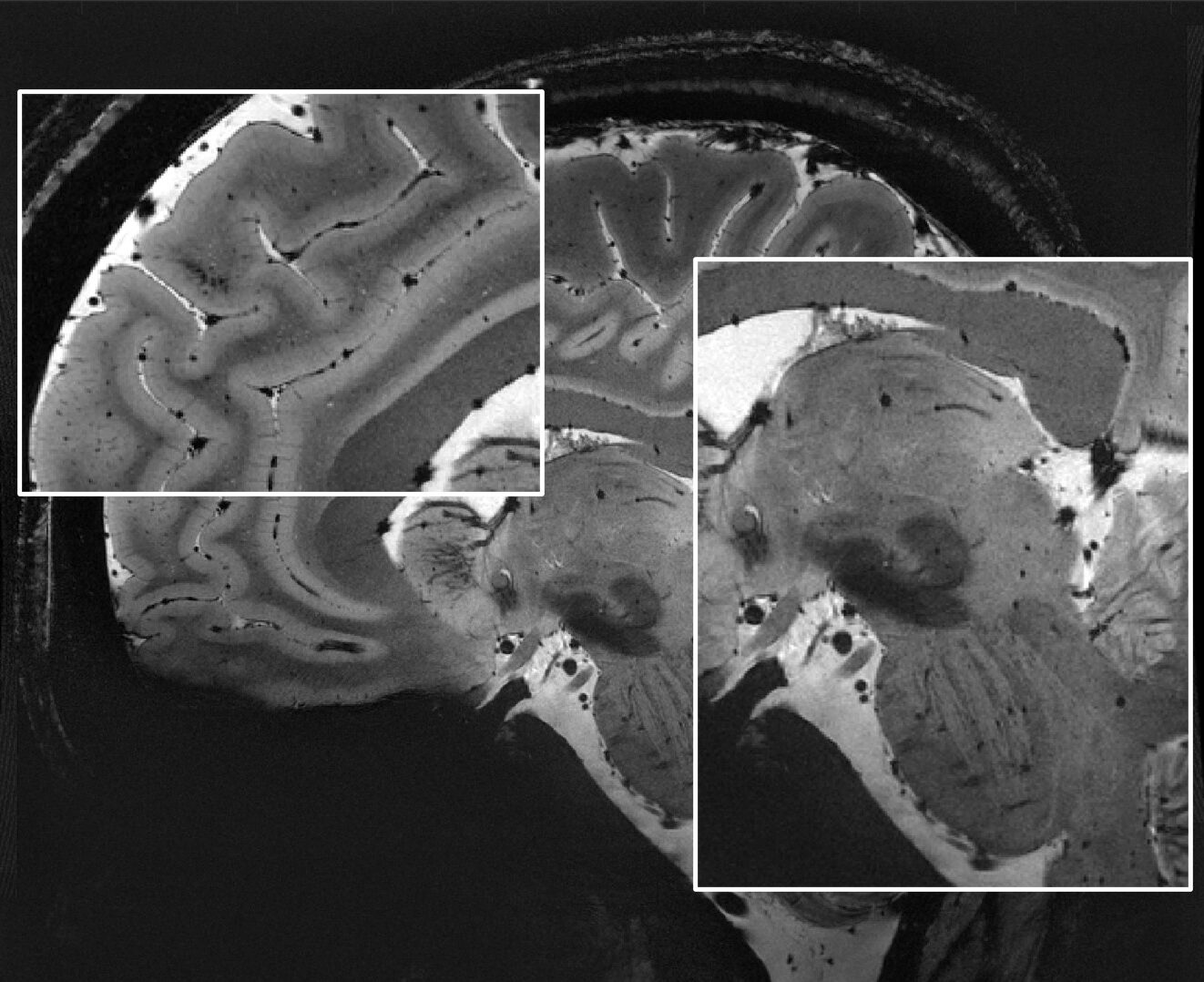
Courtesy: IBT ETH Zurich and University of Zurich
Explore new sequences for cutting-edge fMRI research
The prevalent single-shot EPI acquisition is limited in the range of physiological effects it can capture. The inherently long echo time needed for a high resolution fMRI image prohibits the detection of BOLD activations with short T2* relaxation times. This leads to a failure in identifying the full constellation of neural responses involved in a cognitive task. The NeuroCam allows the use of MR acquisitions such as spirals which can be optimized for sensitivity to the full range of BOLD responses. This freedom in sequence design also benefits the exploration of other physiological effects (e.g. perfusion).