Clip-on Camera™
Flexible sensor platform to monitor existing system performance or to integrate with new hardware.
- MR image acquisitions are corrupted due to field fluctuations from system and physiological sources
- MR system performance can change over long scan sessions, image quality will change along with it
- Living, breathing humans are often the scan subjects, and they influence the encoding fields through different types of motion
Dynamic field fluctuations that are present during MR image acquisitions lead to reduced image quality and image artifacts. There is a wide variety of sources for these fluctuations that impact the field over different timescales. Short term field fluctuations, from microseconds to seconds, occur as a result of eddy currents from encoding gradients, patient motion interacting with the main field of the scanner, mechanical vibration, and other sources. Long term fluctuations, with characteristic times from seconds to minutes to hours can result from gradient heating, with characteristic mechanical resonances changing in response to epoxy stiffness in the coil itself. Longer term changes can also be caused by main field drift due to energy imparted in the system by the pulsing gradient field.
In vivo field monitoring directly measures non-idealities in the encoding field arising from both long- and short-term fluctuations. Image data can then be corrected for these fluctuations, restoring image fidelity and SNR.
What would it mean for your imaging if you could improve image SNR? Field monitoring can compliment existing sequences and readout trajectories and can be used to enable new sequences and readouts, some of which have intrinsically better signal properties than traditional acquisition strategies.
Know your experiment using the Clip-on Camera
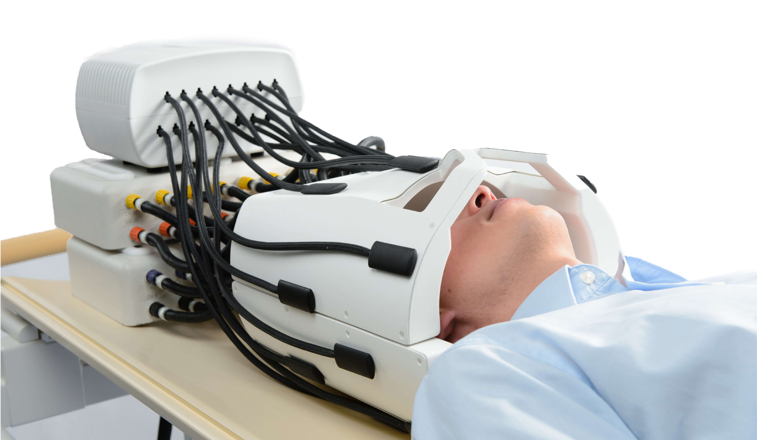
The Clip-on Camera is the add-on measurement system for your existing coil that allows you to fully characterize the dynamic encoding fields while scanning.
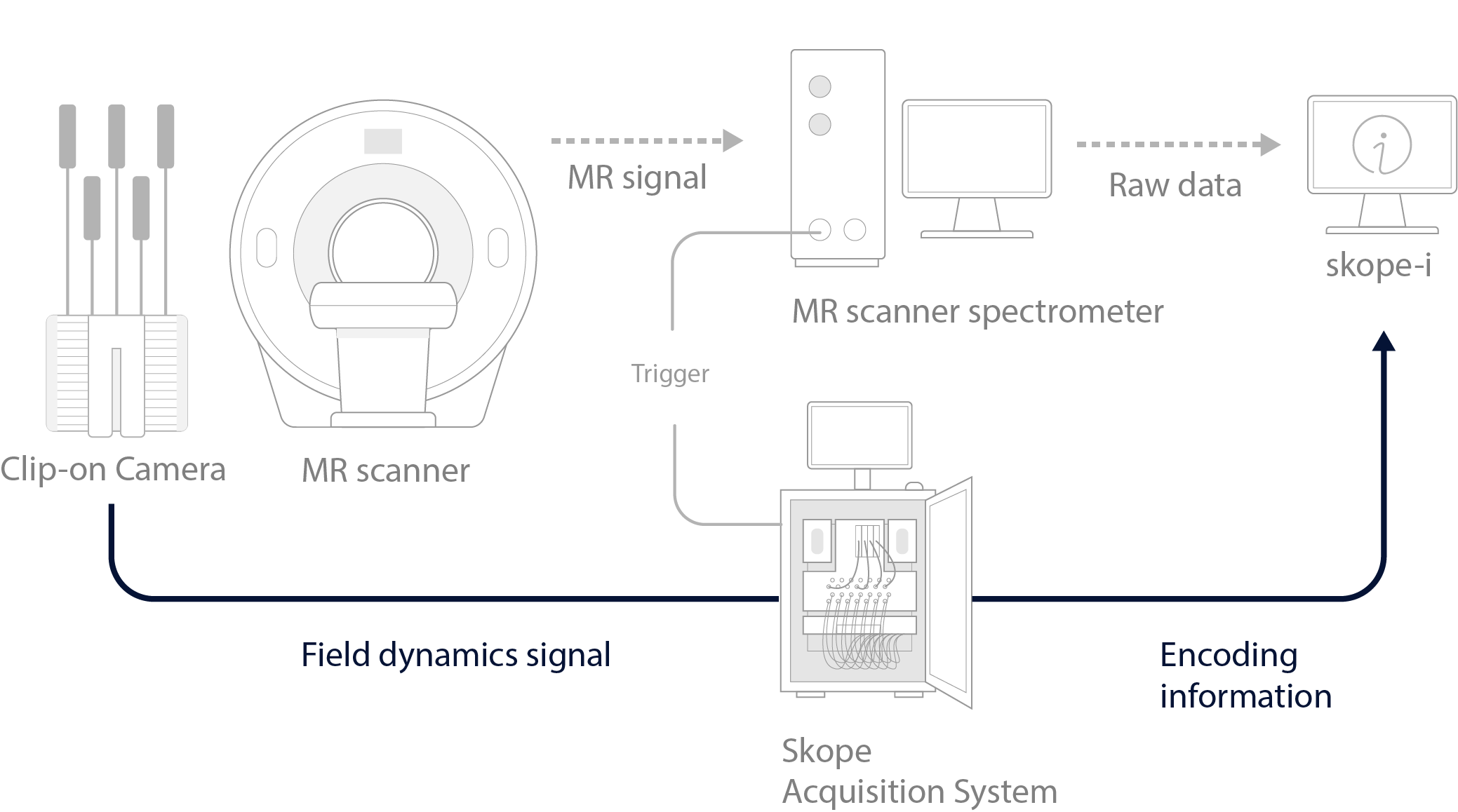
Monitor and record spatial encoding waveforms in addition to dynamic fluctuations from physiological and scanner sources. The Clip-on Camera consists of 16 19F NMR field probes that attach to existing coils or integrate into your new coil design. Using an existing coil allows field monitoring to be integrated into an existing imaging workflow, especially when combined with skope-i, the Skope image reconstruction software package.
For MR technology labs who want to fully leverage their imaging capabilities with concurrent field monitoring, the Clip-on Camera provides direct insight to the actual encoding fields.
Reconstruct accelerated images without geometric distortion
How would your research improve if your images were free of geometric distortion? Geometric distortion is commonly seen in diffusion imaging and is a result of eddy currents from the diffusion encoding gradients influencing the spatial encoding of the imaging readout module. Commonly, the imaging readout module for diffusion acquisition is EPI, or Echo-planar imaging. Each diffusion direction has a characteristic stretch and skew when the image encoding utilizes the EPI readout. Directly measuring and accounting for these in reconstruction allows you to start your analysis with images accurately reflecting the underlying anatomy without having to apply additional corrections methods.
Leverage Neuroscience research with higher image and temporal SNR
Neuroscience research asks fundamental questions about human cognition and behavior. To answer these questions, researchers need imaging methods which increase the SNR of image datasets, allowing for subtle neurological processes to be detected and differences between study groups to be reliably differentiated.
The image above shows how diffusion encoding gradients can impact image geometry with an EPI readout. Six encoding directions and a b=0 image were acquired. The left panel of the image shows the scanner reconstruction and exhibits stretch and skew caused by eddy currents from the diffusion encoding gradients. The right panel shows those same images reconstructed using field evolution data captured by the Clip-on Camera and reconstructed in skope-i. Reconstruction that accounts for field evolution removes geometric distortion from images.
Being able to correct for dynamic field fluctuations, as measured concurrently with the image acquisition, allows to greatly improve image quality. But you can even go one step further, introducing novel acquisition schemes. Since the actual trajectory is measured, otherwise challenging acquisitions such as non-uniform EPIs or spirals, can be used without the need for cumbersome calibrations.
Monitor image encoding in vivo at UHF
Ultra-high field (UHF) imaging has many potential advantages for neuroscience research. UHF promises increased spatial resolution and increased SNR. However, it comes with a wide variety of challenges as well.
As field strength and spatial resolution increases, the potential for image corruption from magnetic field fluctuations caused by subject breathing and cardiac motion outside the imaging field of view increases. Since these field fluctuations during the imaging experiment can be measured in real time, we know the impact on the spatial encoding of the image.
We then use this information to produce the best images possible through either retrospectively compensating for dynamic field fluctuations from the data or as a source of data to update the spatial encoding parameters of the scan in real-time.
 |
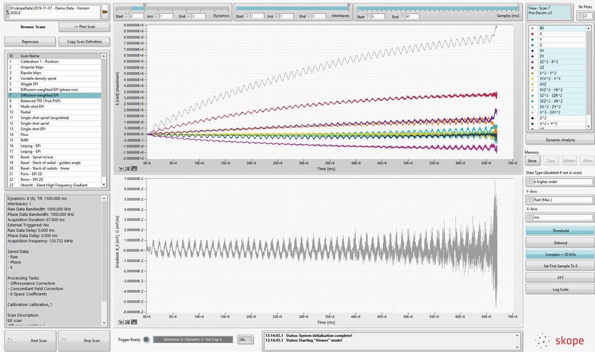
The skope-fm software controls the acquisition and processing of the field data and allows for a fast and easy visualization.
|
 |
 |
“Cranberry” EditionThe Clip-On Camera “Cranberry” Edition comes fully configured to operate in your 7T scanner, which improves workflow for configuration and integration. Based on our well-established Clip-On Camera, “Cranberry” delivers our neuroimaging solution right at the doorstep of your 7T scanner. |
 |
Image production software supporting expanded encoding model algebraic reconstruction
Data conditioning for Image Reconstruction: (Included in skope-i) In order to apply your own reconstruction you will need to resample trajectory information to be on the same time-base as the i-q pairs coming off the scanner. This toolbox provides the tools to measure and account for delays between the MR system and the Skope Acquisition System. |
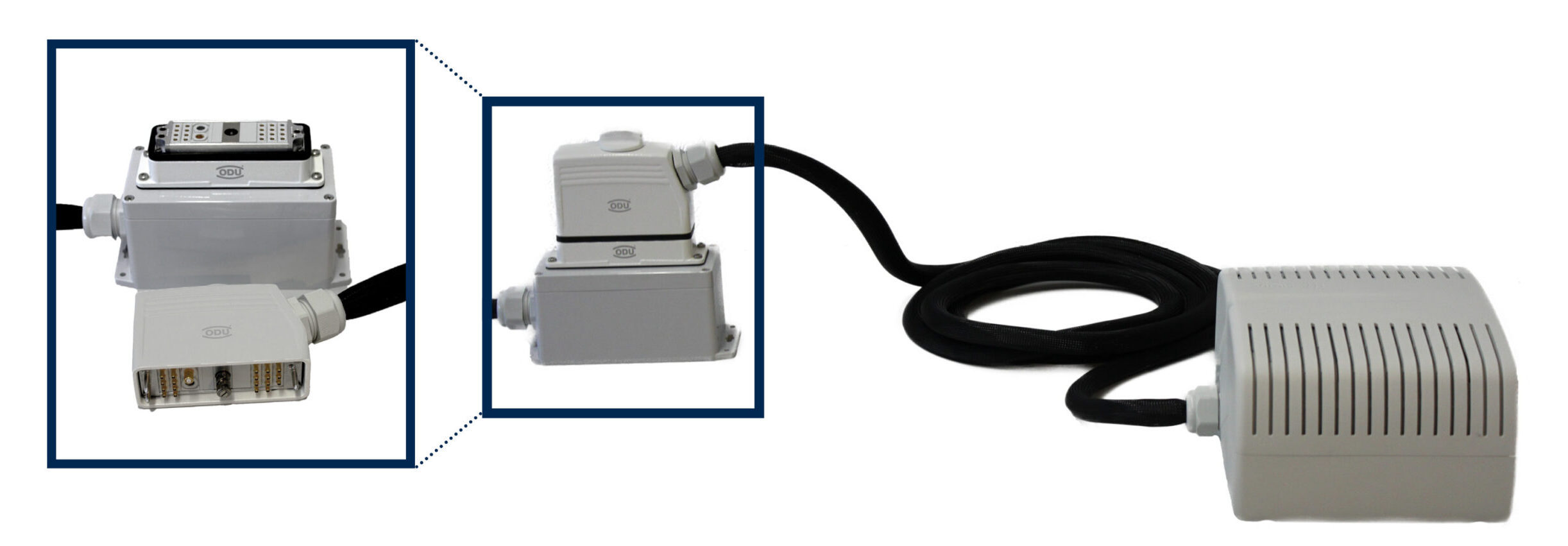
Connect your Skope Camera in one touch and save time when connecting your Skope Camera to the Camera Acquisition System.
Connect Clip-on Camera probes more easily to the front end box with click-in micro-coaxial connectors, saving time when setting up your experiment.