Ultra-High-Field Imaging
UHF imaging has many advantages for neuroscience research, though it comes with many additional challenges. Field monitoring can help you achieve images with high SNR and accurate geometry that advance your UHF research goals.
Ultra-high field imaging (UHF) promises increases in image SNR, resolution, and contrast that have the potential to dramatically improve both research and clinical applications. UHF imaging can allow researchers and clinicians to detect smaller tumors, multiple sclerosis lesions, and epileptic foci, enabling earlier treatment and providing better outcomes. Increases in contrast and SNR have the potential to improve neuroscience research through the more reliable detection of neurological activation and smaller fiber tracts.
However, taking advantage of these promised improvements requires significant technical expertise, as UHF imaging comes with challenges:
- Artifacts from physiological motion (breathing, cardiac cycle) outside the imaging field-of-view become more pronounced at the high spatial resolution UHF imaging promises
- B0 fluctuations and eddy currents can be less ideal at higher fields strengths as every system component is more highly stressed
- Shading artifacts are more common due to localized transmit fields generally required for use at 7T and above
Field monitoring of UHF imaging permits direct measurement of the dynamic field within the UHF scanner. Image reconstructions informed by field monitoring correct for encoding errors. Using field monitoring, you can easily address field fluctuation due to physiological motion, B0 dynamics, and image-encoding related quality issues, allowing you to focus more on your research and UHF developments.
A particular promise of UHF imaging is that of trading the higher intrinsic signal to noise offered by the higher static field for higher spatial resolution. However, higher resolution images are more susceptible to field perturbations from physiological motion due to cardiac and respiratory cycles. Subject motion, even subject motion outside the field of view, can cause visible image artifacts. These artifacts are a result of the subject’s body coupling to the scanner’s magnetic field. When the subject moves, the field changes. If the subject moves during a readout train, the field change can result in blurring, shading, and ghosting artifacts. Below, we see a measurement of the field change due to two different types of motion in a 7T human MR system, measured with an NMR sensor such as a Clip-on Camera.
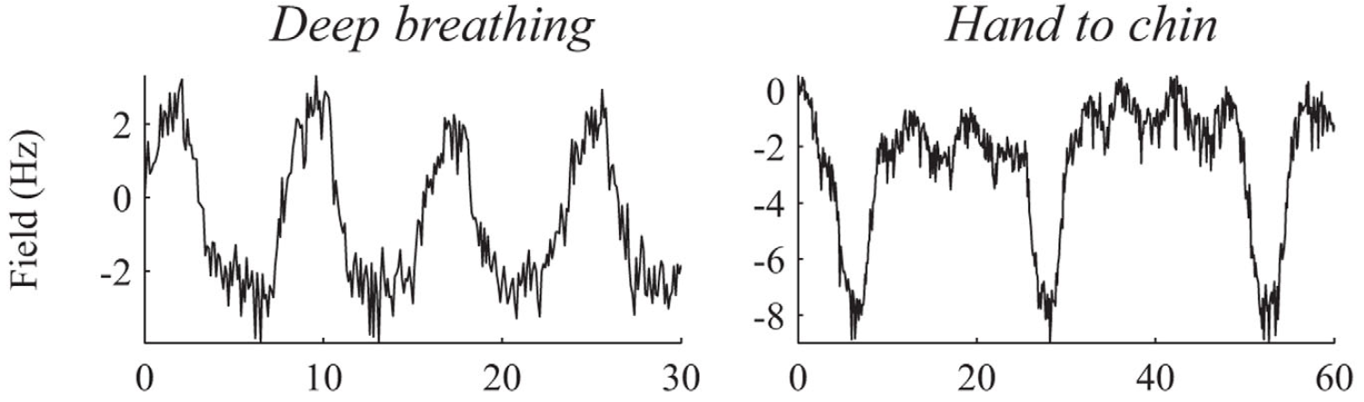
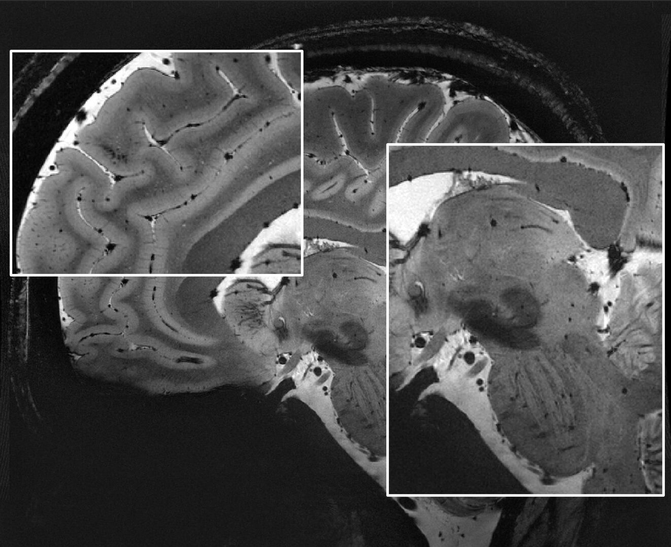
Field monitoring directly measures the field of the scanner during image acquisition, capturing physiologically induced field fluctuations. The graphs above show that deep breathing and hand to chin motion can cause field fluctuations on the order of several Hz. These fluctuations can have an impact on image quality. Direct measurements of the field obtained through field monitoring can be used to correct image encoding, reducing the appearance of artifacts and improving image quality. The image below shows the effect of reconstructing images with and without field monitoring during a deep breathing exercise. Left image is reconstructed with field monitoring, note the reduction of shading artifacts.
Image shading at ultra-high field: not just B1+!
Image shading (aberrant fluctuations in image intensity throughout the image) is commonly seen in UHF images. The transmit (B1+) RF field is commonly assigned as the culprit and indeed it is a main contributor to the artefactual shading. However, it is not the only source of shading. Non-ideal image encoding and physiological motion from between data acquisition windows can also contribute to shading. Field monitoring provides two ways of dealing with this motion. As we saw above from Vannesjo et al., we could use the knowledge of the physiologically induced encoding fields to correct image shading in the reconstruction process.
Alternatively spatiotemporal field measurement was used by Wyss et al. to dynamically update the spherical-harmonic resistive shim currents, allowing them to compensate for fluctuations in the B0 field caused by patient breathing, and even for the field effects of the subject touching their chin with their hand. They applied this to T2* weighted imaging and realized improvements in image quality. Subsequently this improved image quality applied to T2* relaxation mapping resulted in distinct improvements in the quality of their quantitative maps.
Higher resolution imaging allows researchers to see smaller details in the underlying physiology. Ultra-high field (7T +) holds multiple promises for improving image resolution by starting off at a higher image signal-to-noise. Spatiotemporal field measurements improve these images by allowing you to correct for spatial encoding imperfections. In order to acquire a high spatial resolution image, one must cover the relevant portions of acquisition space (k-space) quickly before the signal dephases away due to T2 or T2*. The longer one spends reading out, the more susceptible one is to any source of nuisance phase imparted by unaccounted for encoding fields, which cause the spatial encoding to deviate away form nominal. At higher and higher nominal spatial resolution, even small deviations from nominal encode can degrade your actual spatial resolution, and cause all that extra time and magnetic field strength to be wasted!
In order to effectively use all that field strength, and to get out to the edges of k-space where the detail lies, we must have an efficient acquisition method which is accurately understood. Spatiotemporal field monitoring at UHF via Dynamic Field Camera or Clip-on Camera is that solution. Combining these solutions with skope-i, our image production software, you now have a complete solution to fully unlock the key to UHF.