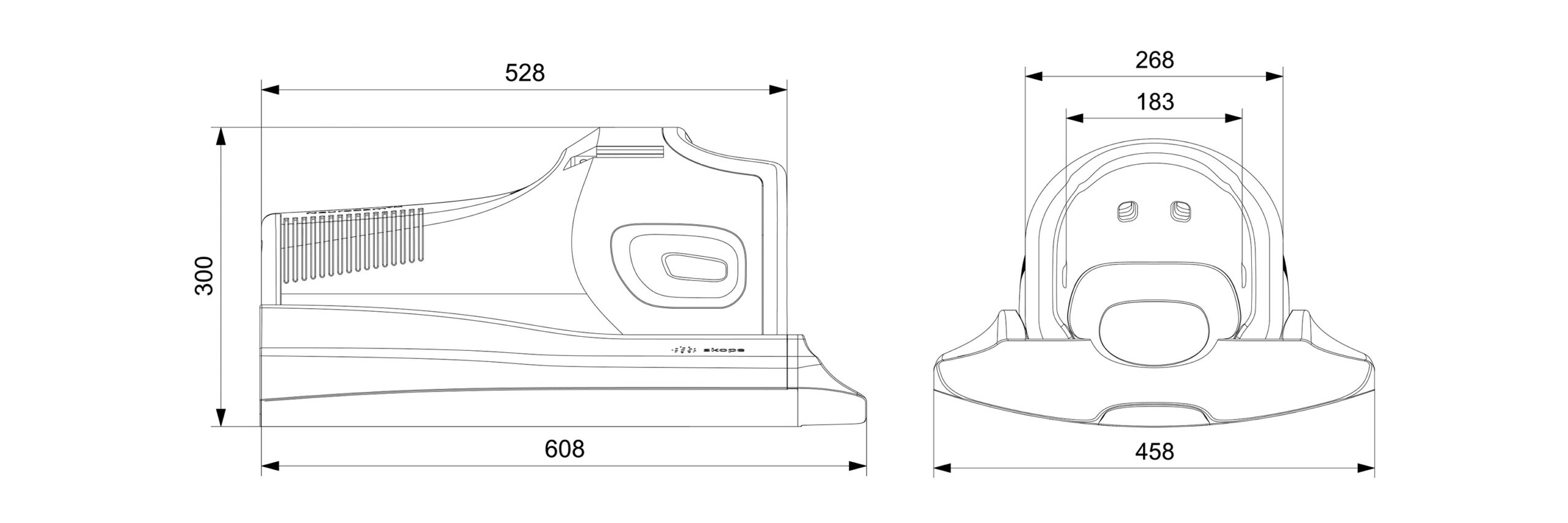
NeuroCam™ 3T
Brain coil with fully integrated field monitoring. Compatible with all 3T MR systems and designed for neuroscientists who want faster scanning and increased spatial resolution via higher base SNR.
- Variability across subjects, scanner types and MRI sequence results in comparability issues for neuroscience
- Encoding field perturbations compromise image quality. These originate from scanner imperfection (eddy currents, etc.), physiological motion (breathing), global image distortions (imperfect B0 shimming), and susceptibility artifacts (local B0 distortion)
- Spatial and temporal resolution trade-offs in neuroscience applications compromise data quality
Today real world gradient fields are very close to the ideal case, yet, the remaining imperfections still lead to image inaccuracies – sometimes invisible to the eye, sometimes in the image phase only – that are affecting the scientific data analysis.
Neuroscience imaging requires a high precision and repeatability to ensure comparable and reproducible results across scan session, subjects, scanners, or sites. Non-reproducible imaging can have many sources – patient motion, patient induced field changes, scanner software version, scanner state, slice positioning – the list can be endless! Given all these sources of error, the imaging analysis pipeline is tasked with the heavy burden of somehow harmonizing all these potential sources of variability and pulling out a significant result which informs the research question.
The standard acquisition time sometimes does not allow to reach higher resolution images due to the increase of acquisition time that will be needed and that is not sustainable for the patient, the scanning time and the scanning budget. Acquiring images with the NeuroCam™ 3T will allow your center to work with your standard acquisition time whilst trading the up to 80% SNR increase for increasing effective in-plane resolution in your images by up to 35% (Lee et al., 2020).
Brain coil with integrated field monitoring for accurate neuroimaging
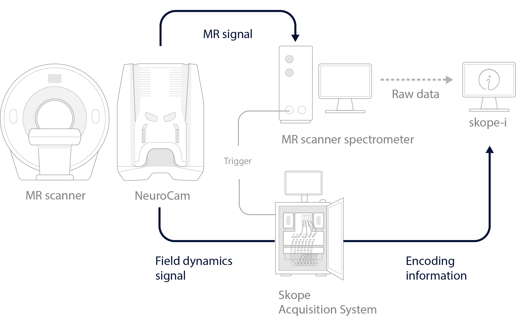
Additional to standard image acquisition, you now can acquire and “monitor” the encoding information of the (imperfect) gradient fields. The NeuroCam™ 3T allows you to make an image with excellent receive performance whilst tracking the influence of the MR system and of the patient that perturb your image formation.
For neuroscientists who need the most accurate, robust, and reproducible MRI scan results, the NeuroCam™ 3T facilitates image quality improvements through active measurement and correction of dynamic field inhomogeneity.
Correction for encoding field deviations caused by the system and the volunteers’ anatomy and movements allows to assess neuro-data in a truly objective manner which will improve structural images, DWI and fMRI data.
Single-shot acquisitions greatly profit from field monitoring and are inherently robust against motion artifacts.

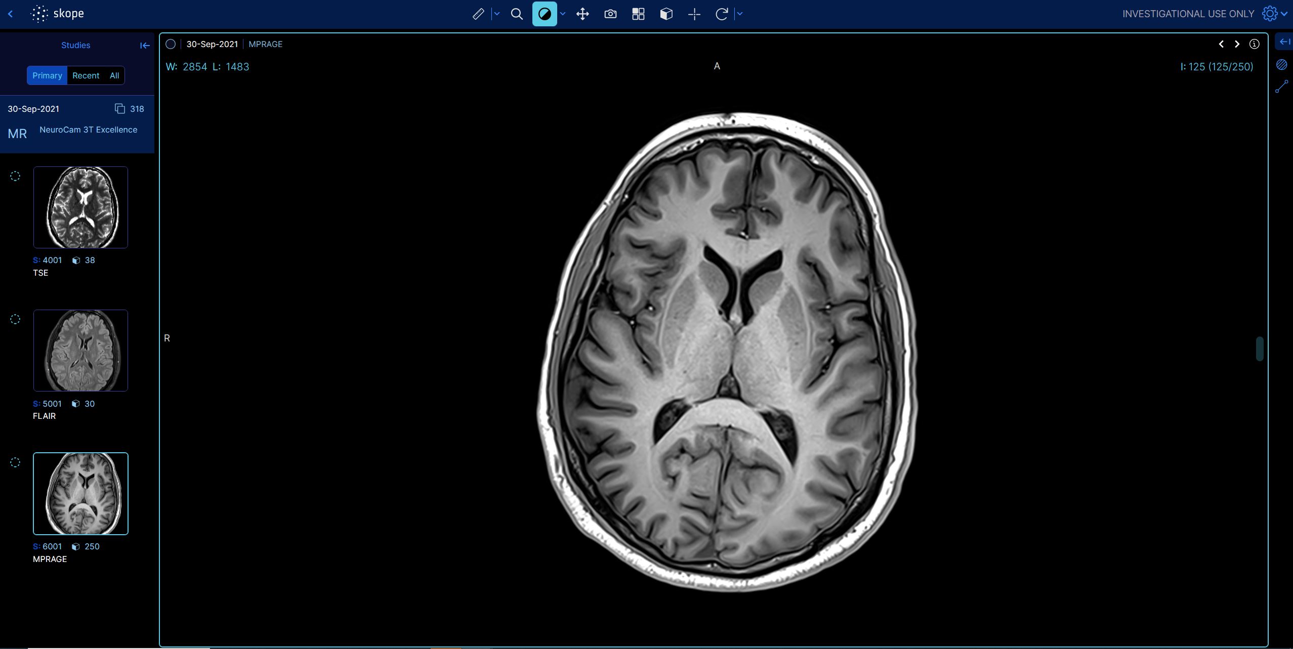
DICOM Viewer
 |
Higher spatial and temporal resolution with the NeuroCam™ 3T
Using the NeuroCam™ 3T with its field monitoring capabilities, spiral imaging increases SNR by 36-88% compared with standard EPI variants. This can be traded either for higher spatial or higher temporal resolution – increasing the significance of findings or reducing the overall exam time. For example, an 80% increase in SNR can be used to reduce each edge of a voxel by 20%, maintaining the same acquisition time.
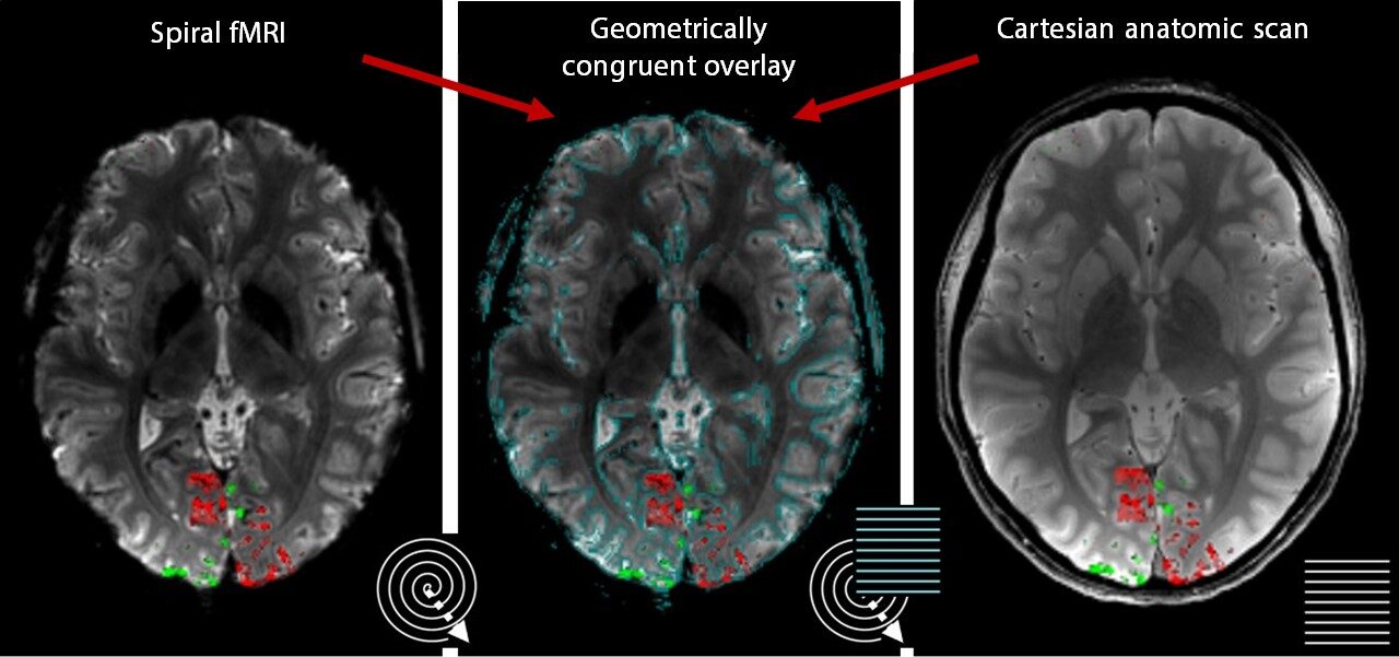
Improved geometrical congruence
The geometrical misalignment between individuals in an fMRI study leads to erroneous anatomical allocation of the group-level BOLD signal or even a failure to detect a population effect. Moreover, multi-modal imaging studies can fail due to anatomical inconsistency between different contrasts (e.g., fMRI and DWI). Measuring the imperfect encoding fields, the geometrical congruence can be recovered, thus enabling reliable group-level analyses and multi-modal imaging studies.
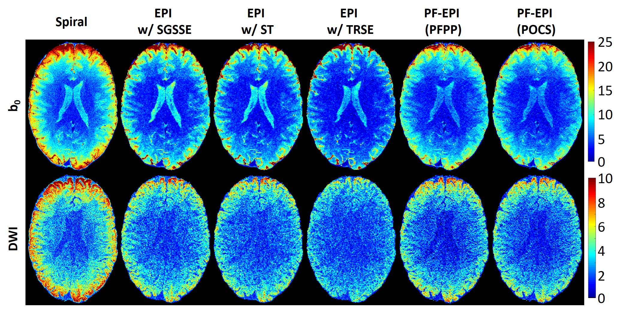
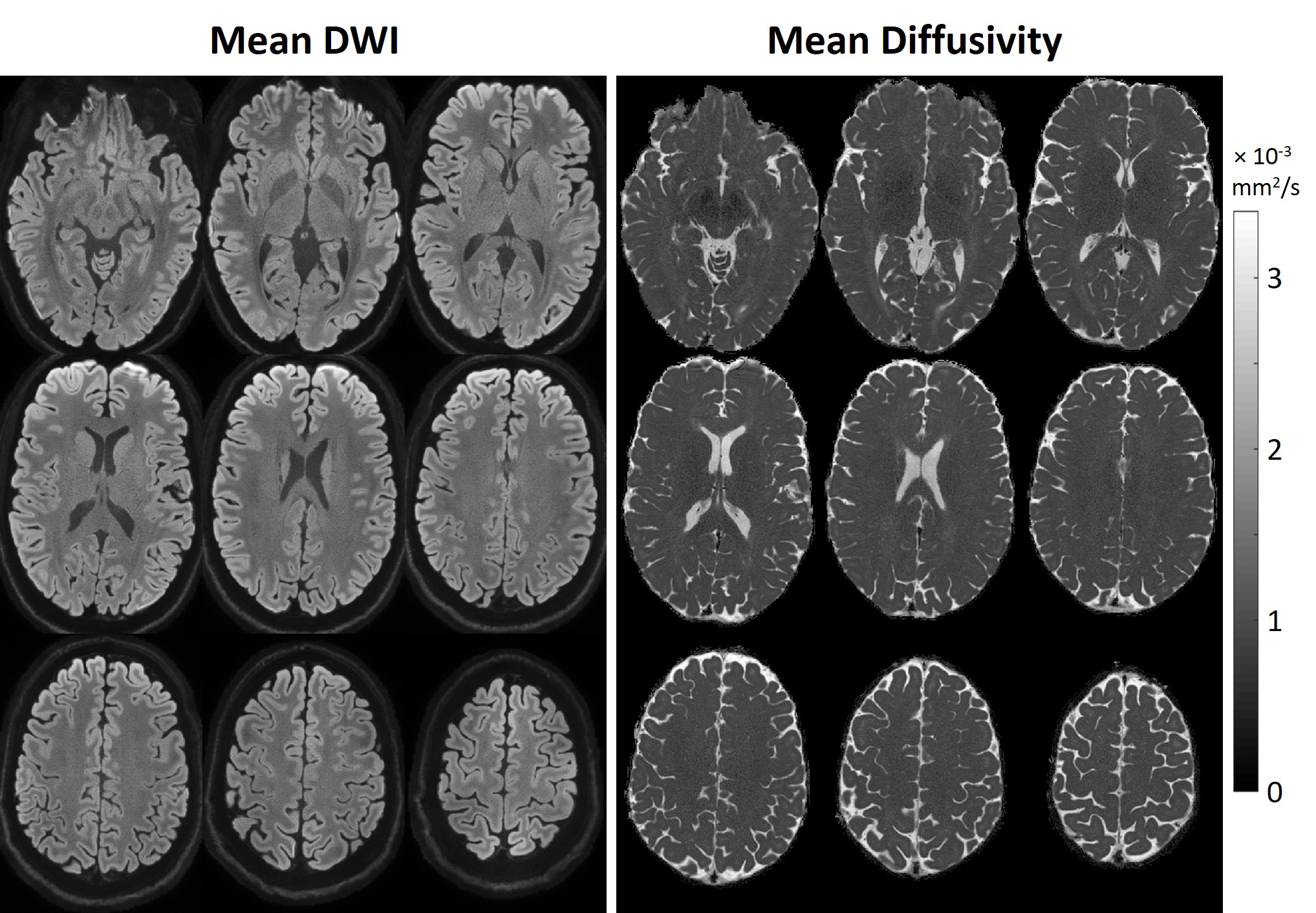
Improved effective image resolution without affecting acquisition time
With the NeuroCam™ 3T, the effective resolution can be improved without affecting acquisition time. The gain in SNR by up to 80% (Lee et al., 2020) can be traded for higher spatial resolution without affecting the acquisition time. Below are examples of diffusion-weighted images acquired with the NeuroCam™ 3T.
 |
skope-i image reconstruction software that will integrate the monitoring information collected by the NeuroCam for production of the highest SNR images. |
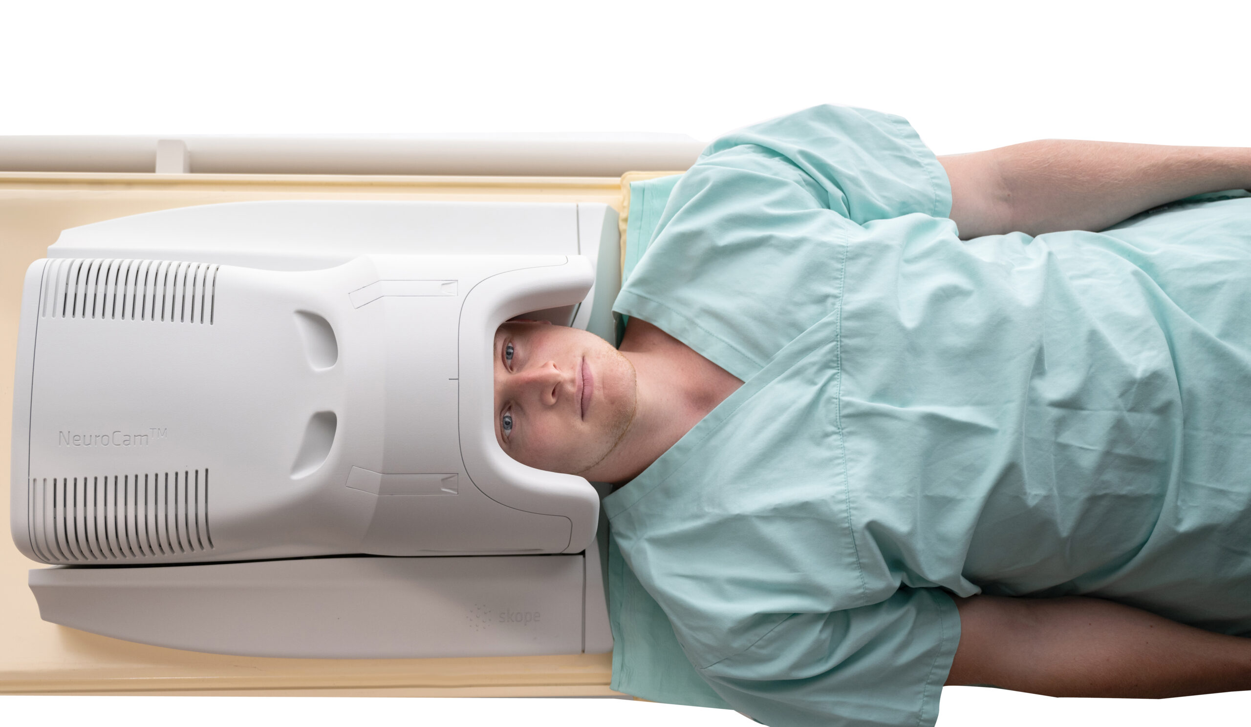
The open coil towards the face allows multimodal imaging with additional devices such as eye tracking, mirror fixing, open field of view for back vision satisfying needs from different cognitive neuroscience applications.